
What is the difference between 2d, 3D, 4d & 5d
Top of the line technology
-

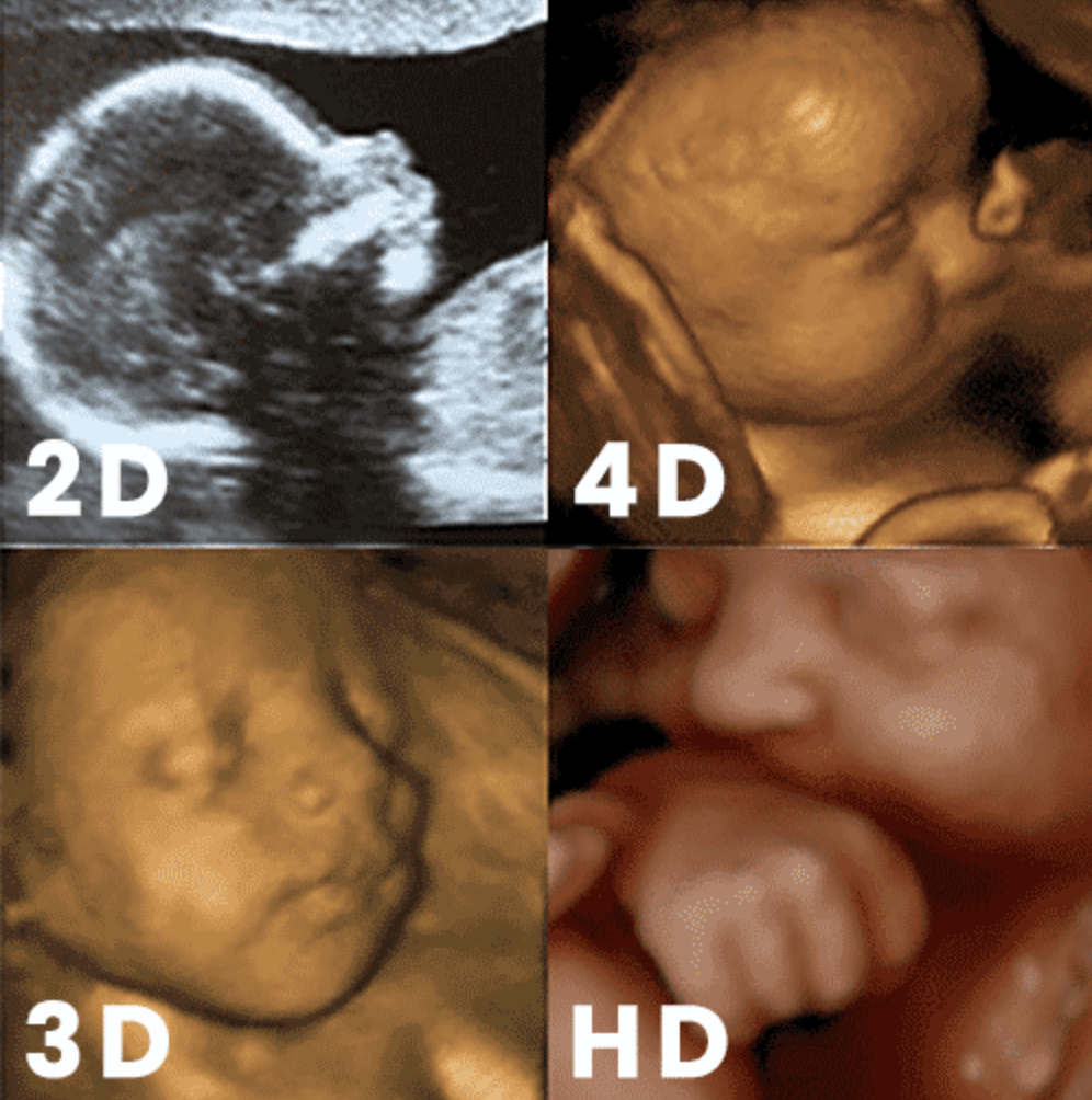
2D
As the most common form of ultrasound, a 2D ultrasound creates a black and white image that shows the skeletal structure of the baby and makes the internal organs visible. The 2D ultrasound is most commonly used to diagnosis the health of the baby. As the name implies, all 2D images are flat and have no depth to them.
-

3D
Much like 2D ultrasounds, a 3D ultrasound uses soundwaves to develop an image of the baby inside of your womb. However, unlike the flat image given by a 2D ultrasound, a 3D ultrasound creates the appearance of a three-dimensional image of the baby. This allows the expecting parents to see their baby’s face, rather than just the outline of the face.
-

4D
4D ultrasounds also create a three-dimensional image and provide an even clear image of the baby. The 4D ultrasound is created while capturing a video of your baby. We then scroll through the video and save still images. With 4D ultrasound imaging, you could potentially see your baby smile or yawn in the womb because it compiles hundreds of images into a moving video.
-

5D/HD live
As the most recent advancement in ultrasound technology, HD and HD Live (also called 5D Ultrasound ) ultrasounds allow us to capture even clearer, sharper images. These images are more defined and have better resolution.